1/28/2004   slide 10
Retinal Array
•You somehow construct lines, curves, etc from patterns transmitted by this neuron array
•Visual cortex has cells sensitive to:
–Line orientation: horizontal, vertical, and diagonal,
–Changes in brightness (edge detection)
–Length
–Motion
–Even direction of motion
–and more…
•Movie of retinal excitation (guinea pig) in response to moving bars http://www.snl-e.salk.edu/technology/
•Vision is “noisy”—massive statistical processing apparently takes place
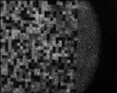
Retinal readout, hundreds of cells of guinea pig retina, Salk Institute
http://www.snl-e.salk.edu/technology/RRS/2003-03-24/source/array-stimulus.tiff.html
Like digicam CCD

Terabytes of data generated by tiny fraction of output of a whole retina. Brain does awesome data processing job.

The goal of the Retina Project is to understand the language the eye uses to send information about the visual world to the brain, to study how a biological neural network (the retina) processes and encodes information. This is a truly interdisciplinary collaborative effort between high energy physicists from SCIPP, and neurobiologists, experts in nanofabrication, and VLSI designers. The retina is a thin tissue which lines the back of the eye. It is a sophisticated pixel detector that converts an input visual image into a set of highly processed electrical signals ("spikes") that travel up the optic nerve to the brain. These spike trains collectively encode and communicate the critical features of the image in a timely manner to the visual cortex. To study this neural code, we are developing an imaging system for large-scale neural activity. This "Retinal Readout System" will allow the simultaneous recording of signals from hundreds of retinal output neurons (the ganglion cells) as a dynamic visual image is focussed on the input neurons (the photoreceptors). The implementation of this system is based on the silicon microstrip detector technology and expertise we have pioneered and developed in SCIPP to study short-lived particles and CP violation, and to search for the Higgs Boson, in high energy physics experiments. In the neurobiology experiment, live retinal tissue is placed in a chamber on top of an array of microscopic electrodes fabricated on a glass substrate. The chamber is filled with physiological saline solution; retinal tissue superfused in this manner can be kept alive for several hours. A dynamic image on a computer display is focussed on the photoreceptors. In response to this image, the ganglion cells generate spikes which are picked up as extracellular signals by the electrodes. These signals are amplified, filtered, digitized and recorded. The spike trains from the hundreds of neurons detected can then be correlated in space and time among themselves and with the visual image to study the neural code at the retina/brain interface.
http://www.snl-e.salk.edu/technology/